|
|
|
|
|
Aiming for human induced pluripotent stem cell (iPSC) production for clinical use
3D culture with atelocollagen ー Coating and Microcarriers ー
|
|
|
Since the establishment of mouse and human iPSCs was reported in 2006 and 2007, respectively, research on iPSCs has been continuously progressing. Various approaches to the clinical applications of iPSCs are currently in progress. One such approach is the “my iPS® Project,” which was promoted by the Kyoto University iPS Cell Research Foundation.
In this article, we introduce a paper on the establishment of iPSCs in a 3D culture environment and the differentiation induction process related to the my iPS® Project.
|
|
Work processes in cell therapy using autologous iPSCs
|
|
|
|
Many may think that cell therapy, especially those using autologous cells, is an expensive treatment that requires costly cell production. The my iPS® project aims to provide autologous iPSCs to research institutions and companies at a cost of around 1 million yen by constructing a closed automated culture system and its 3D culture process, thereby reducing the cost of establishment, proliferation, and the induction of iPSC differentiation.
|
|
|
|
Atelocollagen for the maintenance of undifferentiated iPS cells in culture
|
|
|
|
|
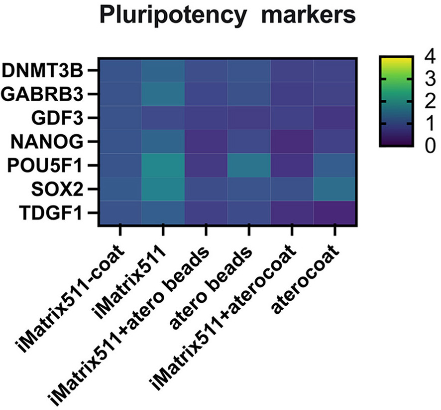
When the iMatrix-511-coated plate was used as a control group, the expression of pluripotency markers did not significantly fluctuate, and the undifferentiated state of the iPSCs was maintained in both the atelocollagen-coated plate and atelocollagen beads (collagen microspheres) groups.
Another experiment indicated that the atelocollagen groups did not affect the endodermal and ectodermal differentiation abilities. The microsphere group was the only group in which the expression level of T, a mesoderm marker, fluctuated more than 4-fold; however, it was confirmed that the increased expression could be suppressed when used in combination with iMatrix-containing medium.
|
|
|
|
Atelocollagen promoted the formation of iPSC filopodia
|
|
|
|
|
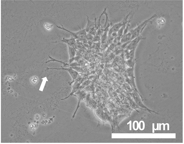
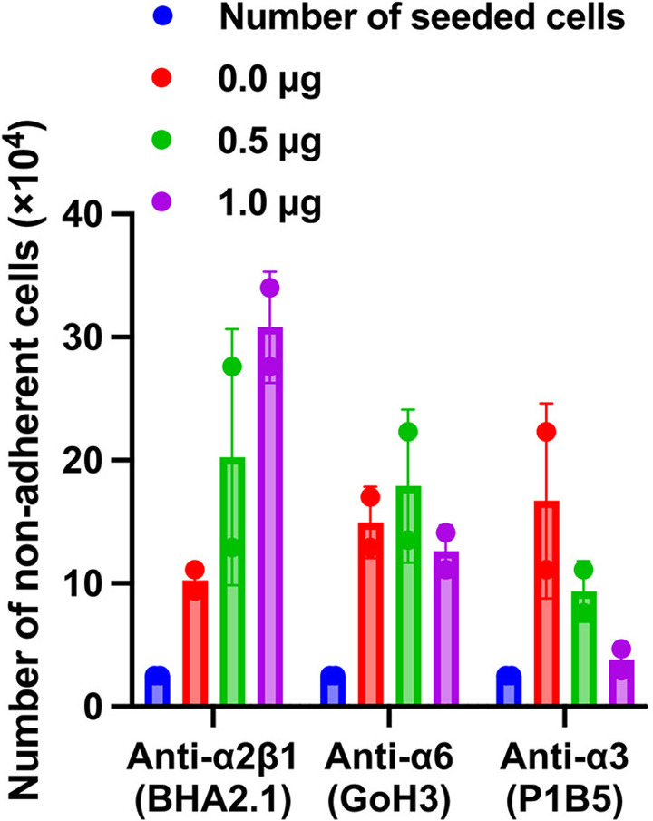
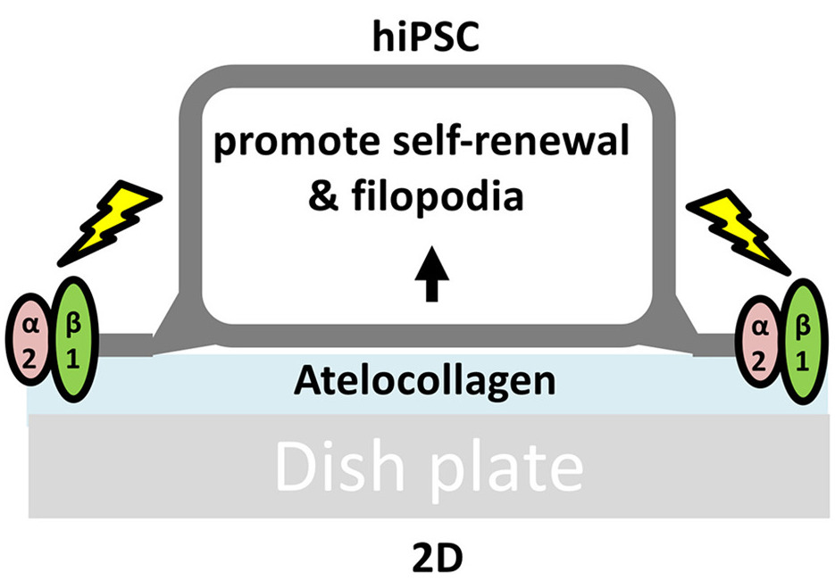
Meanwhile, when iPSCs were cultured on atelocollagen-coated plates, the extension of extremely long filopodia was observed. When inhibitors of and antibodies against integrin α2β1, which is a collagen receptor, were added, the formation of filopodia was suppressed and the number of iPSCs that did not adhere to the plate increased. Thus, it was suggested that adhesion to atelocollagen via integrin α2β1 promoted the self-renewal of iPSCs.
|
|
|
|
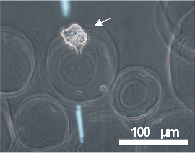
Establishing iPSCs on microspheres
|
|
|
|
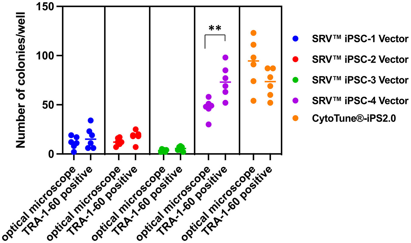
Next, stealth RNA vectors optimized for 2D culture were investigated to determine whether they could also function in a 3D culture environment for the establishment of iPSCs. As a result, iPSC colony formation was confirmed using both optical and fluorescence microscopes to observe TRA-1-60-positive cells.
|
|
|
|
|
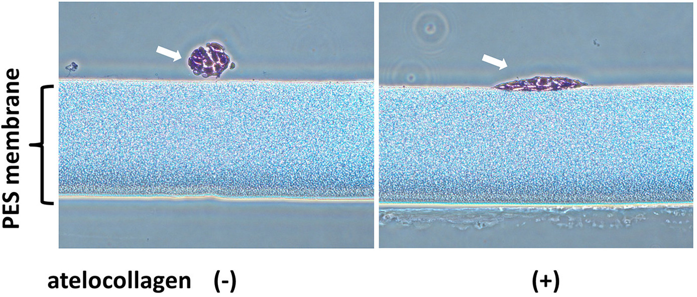
Atelocollagen improved the adhesion of iPSCs to hollow fiber membranes
|
|
|
|
|
Finally, the combined use of the closed automated culture device and atelocollagen was evaluated. iPSCs do not adhere to polyethersulfone (PES) hollow fiber membranes; however, they adhered to the atelocollagen-coated device, and the undifferentiated state was strengthened.
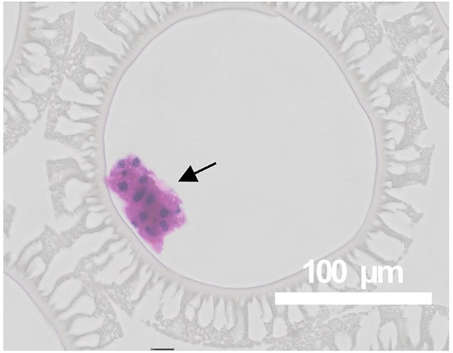
In addition, when iPSCs seeded on microspheres were cultured in a closed vessel and differentiation was induced, they exhibited the ability to differentiate into three germ layers. This suggests that iPSCs can be used for the induction of cellular differentiation for therapeutic use as well as for the establishment of iPSCs for clinical use.
Source:Mol Ther Methods Clin Dev. 2024 Jul 20;32(3):101302.
Created by modifying figure 1-4. ©Nakashima Y., et al. 2024. Licensed under CC BY 4.0
(https://creativecommons.org/licenses/by/4.0/).
|
|
|
|
Two atelocollagen products used in the aforementioned paper
|
|
Atelocollagen acidic solution I-PC 5mg/mL
|
|
|
|
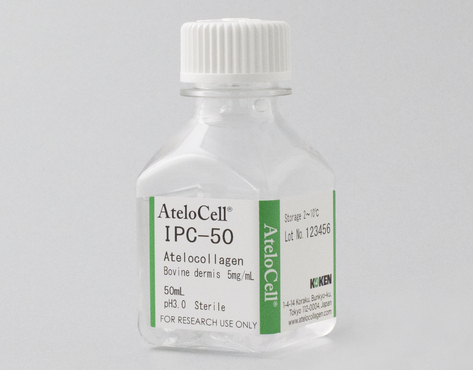
Atelocollagen acidic solution I-PC 5mg/mL(Code: IPC-50)
This product was used to coat the plates, where the iPSCs’ undifferentiated state was maintained and differentiation was induced. It was also used to coat hollow fiber membranes and other components of the closed automated culture devices.
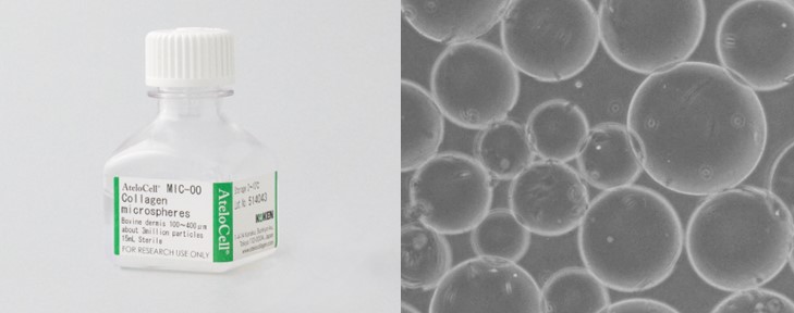
Collagen microspheres (Code: MIC-00)
This product was used to coat the plates where iPSCs were established and induced to differentiate and to coat the closed vessels where iPSC differentiation was induced.
|
|
|
|
|
|
Exhibition information
We plan to participate in two exhibitions in March 2025. Please visit us at the venue to see our various atelocollagen products in person.
1. APPW2025(Joint Meeting of the Japanese Association of Anatomists, the Physiological Society of Japan, and the Japanese Pharmacological Society)
Dates: March 17 (Monday) to March 19 (Wednesday) 2025
Venue: Makuhari Messe, International Exhibition Hall No. 8
Booth:13
2. The 24th Congress of the Japanese Society for Regenerative Medicine
Dates:March 20 (Thursday) to March 22 (Saturday) 2025
Venue:PACIFICO Yokohama North
Booth:B061
|
|
|
|
|
|

.jpg)