Show product listProduct list
- 3D Ready Atelocollagen
- Atelocollagen/Native Collagen Acidic Solutions
- Atelocollagen Neutral Solutions
- 3D Honeycomb Boosted
- Atelocollagen, Honeycomb Sponge
- Atelocollagen Sponge, MIGHTY
- Atelocollagen Coated β-TCP Scaffold
- Atelocollagen Microspheres
- Collagen Sponge for 35 mm Culture Dish
- FibColl® Atelocollagen Inserts 24
- Atelocollagen, Permeable Membrane
- Type II Collagen
- Atelocollagen Powder, Membrane, Sponge
FibColl® Atelocollagen Inserts 24
Outline
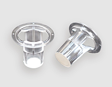
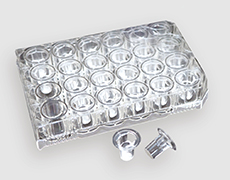
FibColl® Atelocollagen Insert 24 is a novel cell culture insert that uses a membrane entirely made from atelocollagen. Unlike other collagen-coated plastic membranes, FibColl® pores are not clogged, and the fiber structure of atelocollagen enables cell culture in an in vivo mimicking environment. The microporous structure between atelocollagen fibrils allows the permeation of molecules even over 600 kDa, making it suitable for barrier function assessment and co-culture models.
Applications
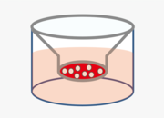
Culture on the membrane
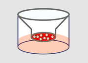
Air-liquid interface culture
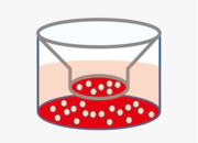
Co-culture

Cell sheet transplantation
Features
- Membranes made from atelocollagen are permeable to molecules with molecular weight even higher than 600 kDa.
- A barrier function assessment model can be developed by tight junction formation of epithelial cells.
- FibColl® is an easy-to-use hanging cell culture insert considering medium exchange and membrane recovery after culture.
- The membrane is biodegradable and therefore it can also be used for cell sheet transplantation.
Types of collagen
Bovine hide derived atelocollagen
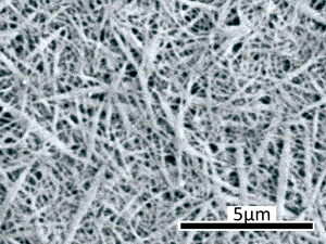
Fibril structure of FibColl® (SEM image)
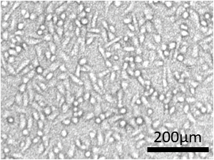
Lung cancer cells cultured on FibColl® for 2 days (optical phase-contrast microscopy image)
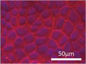
Formation of tight junctions between renal tubular epithelial cells cultured using FibColl® for 2 days (Red: ZO-1, blue: nucleus, fluorescence microscope image).)
| Cat. No. | Product | Packaging | Price |
|---|---|---|---|
| FAI-24 | FibColl® Atelocollagen Inserts 24 | 24 pieces/bag | Visit distributor’s web page |
Storage: Room temperature For research use only.
Click here for frequently asked questions about FibColl® Atelocollagen Inserts
Use case
- Use Case 1
- Use Case 2
- Use Case 3
-
Accelerated maturation of epidermal model using FibColl®
Normal human epidermal keratinocytes (1.8×10⁵ cells) were seeded on the membrane of FibColl® or polyethylene terephthalate cell culture inserts (PET), and cultured submerged for 3 days, followed by air-liquid interface culture for 14 days. Frozen sections of each epidermal model were then observed using HE staining. As a result, a mature stratum corneum was formed on FibColl® as early as day 7 of culture, and further layering of the stratum corneum was confirmed on day 14 (In-house data).
When the expression of each epidermal marker was observed by immunofluorescence staining, maximum expression of CK14, a basal layer marker, was observed near the basal layer with FibColl®.In addition, the staining distinction between CK14 and the spinous layer marker CK10 was clearer than with PET. Furthermore, expression of FLG, a granular layer marker, near the granular layer was prominent (In-house data).
In FibColl® culture, clear expression of CLDN1 and OCLN, components of tight junctions, was confirmed by immunostaining. In addition, TEER measurement showed that FibColl® had a significantly higher TEER value than PET (In-house data).
-
Evaluation of substance permeability and barrier function using FibColl®
①After adding a solution of the evaluation molecule onto the FibColl® membrane, the molecules that migrated under the membrane were quantified. Proteins over 600 kDa also permeated.
② Canine renal tubular epithelial cells (MDCK cells) were cultured on FibColl® for 14 days, and the formation of tight junctions was confirmed by immunofluorescent staining of ZO-1.
③ Transepithelial electrical resistance (TEER) measurements showed electrical resistance.(In-house data)
-
Application of FibColl® in a psoriasis-like epidermal inflammation model
In an epidermal model cultured for 7 days at the air-liquid interface on FibColl®, cytokines were added twice at 24-hour intervals after medium exchange to induce inflammation. Expression of several psoriasis-related genes was found to be upregulated or downregulated, indicating the utility of FibColl® in constructing psoriasis models. Sufficient RNA was extractable from FibColl®(24 well plate size) for qPCR analysis (In-house data).
References
- Culture on the membrane
- Co-culture
- Cell sheet transplantation